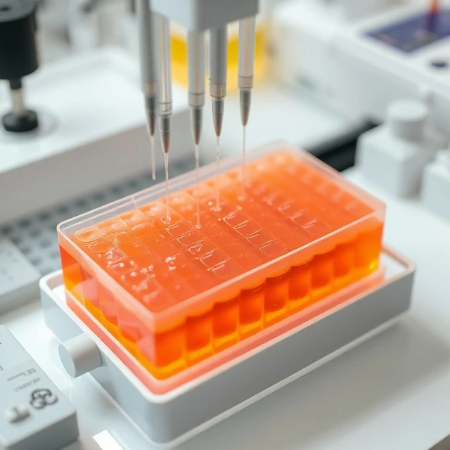
DNA fragments of different lengths can be separated with an electric field. Gel electrophoresis pulls charged molecules through a porous gel matrix; smaller fragments migrate faster than larger ones. The result is a series of bands that reveal the sizes present in a sample.
Agarose gels are used for larger DNA pieces; polyacrylamide gels resolve smaller fragments. DNA is visualized with fluorescent dyes or stains. Comparison to a size standard allows estimation of fragment length. The technique underpins restriction mapping, PCR analysis, and quality control of sequencing libraries.
Although largely replaced by capillary and microfluidic methods for high-throughput work, slab-gel electrophoresis remains a simple, inexpensive teaching and research tool. Proper voltage, gel concentration, and running time determine resolution. The method is foundational in molecular biology laboratories.
- Separates DNA fragments by size
- Uses electric field and porous gel
- Smaller fragments travel farther
- Visualized with fluorescent dyes
Comments
No comments yet. Be the first to share a thought.
Leave a comment