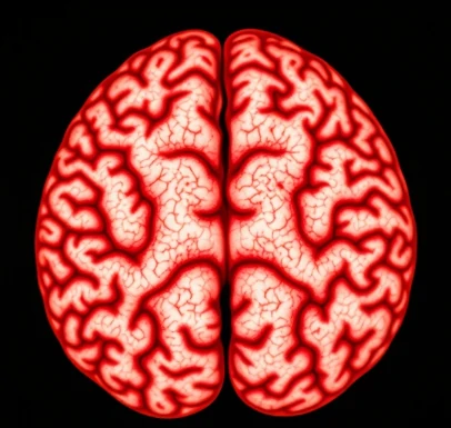
Before function can be understood, structure must be mapped. Neuroanatomy is the discipline that describes the architecture of the nervous system at scales ranging from gross regions to individual synapses.
Classical methods relied on dissection, staining of cell bodies or myelin, and tract-tracing with injected dyes. Modern approaches add immunohistochemistry, genetic labeling, electron microscopy, and noninvasive imaging such as diffusion MRI. The result is a progressively finer atlas of nuclei, cortical areas, fiber tracts, and connectional networks.
Neuroanatomy provides the foundation for interpreting lesions, planning surgery, and designing experiments that target specific circuits. Comparative neuroanatomy reveals both conserved motifs and species-specific specializations. Even as functional studies proliferate, accurate anatomical knowledge remains indispensable; a signal recorded in the wrong place or a stimulation site misidentified can lead research astray.
- Study of nervous-system structure
- Spans macroscopic to ultrastructural scales
- Uses classical and modern labeling methods
- Essential for functional interpretation
Comments
No comments yet. Be the first to share a thought.
Leave a comment