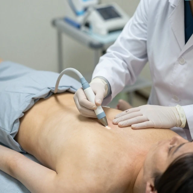
Thoracentesis removes fluid or air from the space between the lungs and the chest wall. That space, the pleural cavity, normally holds only a thin film of fluid. When it fills with excess fluid, called a pleural effusion, breathing becomes difficult. Draining it brings relief and provides a sample for diagnosis.
The procedure is usually done at the bedside or in a clinic. The patient sits upright, leaning forward. The doctor cleans the skin, numbs the area, and inserts a needle or catheter between the ribs. Fluid drains into a container. An ultrasound often guides the needle to avoid the lung and major blood vessels.
Why thoracentesis is performed
- Relieve shortness of breath from a large effusion.
- Test fluid for infection, cancer cells, or heart failure markers.
- Remove air from the pleural space after a collapsed lung.
- Determine the cause of unexplained fluid buildup.
Risks include bleeding, infection, and a collapsed lung, though serious complications are uncommon when ultrasound is used. The fluid itself tells a story. Clear straw-colored fluid suggests heart failure. Cloudy or foul-smelling fluid points to infection. Bloody fluid raises concern for cancer or pulmonary embolism. Thoracentesis is both a treatment and a diagnostic tool, and it can provide rapid relief for patients struggling to breathe.
Comments
No comments yet. Be the first to share a thought.
Leave a comment