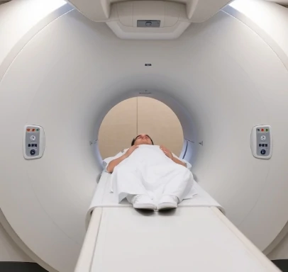
A CT scan, or computed tomography scan, uses a series of X-ray images taken from different angles to create cross-sectional pictures of the brain or body. A computer assembles the images into detailed slices that reveal structure in far greater detail than a single X-ray.
In psychology and neurology the technique is used to detect tumors, bleeding, stroke damage, and structural abnormalities. It is relatively fast and widely available, making it a first-line tool in emergency settings. The patient lies still on a table that slides into a doughnut-shaped scanner; the X-ray tube rotates around the head, collecting data that are reconstructed into images.
CT provides excellent spatial resolution of bone and acute blood, yet it is less sensitive than MRI for soft-tissue differences and involves ionizing radiation. For that reason it is used judiciously, especially in children and in repeated examinations. Contrast agents can be injected to highlight blood vessels or certain lesions.
When a clinician needs a rapid look at brain structure after trauma or sudden symptoms, the CT scan remains a workhorse of modern medicine.
Comments
No comments yet. Be the first to share a thought.
Leave a comment