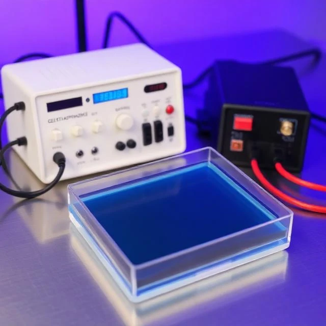
DNA fragments are placed in a gel. An electric current is applied. The fragments migrate through the gel, with smaller fragments moving faster than larger ones. After a few hours, the fragments are separated by size. That is gel electrophoresis, a technique that has been a staple of molecular biology since the 1970s.
The gel is usually agarose for DNA or polyacrylamide for proteins. The gel acts as a molecular sieve. The electric field drives the negatively charged DNA toward the positive electrode. The pore size of the gel determines the range of separation. After electrophoresis, the fragments are visualized with a stain or a fluorescent dye. The result is a pattern of bands, each corresponding to a fragment of a particular size.
Gel electrophoresis is used for many purposes. It checks the size of DNA fragments after PCR or restriction digestion. It separates proteins for Western blotting. It analyzes DNA fingerprints for forensic identification. It diagnoses genetic disorders by detecting abnormal fragment patterns. It is simple, cheap, and informative.
The technique has limitations. It is slow. It is qualitative or semi-quantitative. It cannot resolve very large fragments. But it remains a workhorse of the lab, and it is often the first step in a longer analysis.
Comments
No comments yet. Be the first to share a thought.
Leave a comment