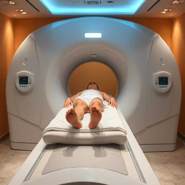
MRI, or magnetic resonance imaging, uses strong magnetic fields and radio waves to produce detailed images of the brain and other soft tissues. It does not rely on ionizing radiation.
The technique detects the behavior of hydrogen nuclei in water molecules when they are placed in a magnetic field and perturbed by radio-frequency pulses. Different tissues return to equilibrium at different rates, generating contrast that reveals anatomical structure with high spatial resolution. Structural MRI is widely used to detect tumors, lesions, developmental anomalies, and the tissue loss associated with neurodegenerative disease.
Functional MRI extends the method by tracking blood-oxygen-level-dependent signals that change with neural activity. The combination of structural and functional imaging has transformed both clinical diagnosis and cognitive neuroscience. Claustrophobia, the presence of certain metal implants, and the cost and availability of scanners remain practical limitations.
When fine anatomical detail of soft tissue is required without X-rays, MRI is the method of choice.
Comments
No comments yet. Be the first to share a thought.
Leave a comment