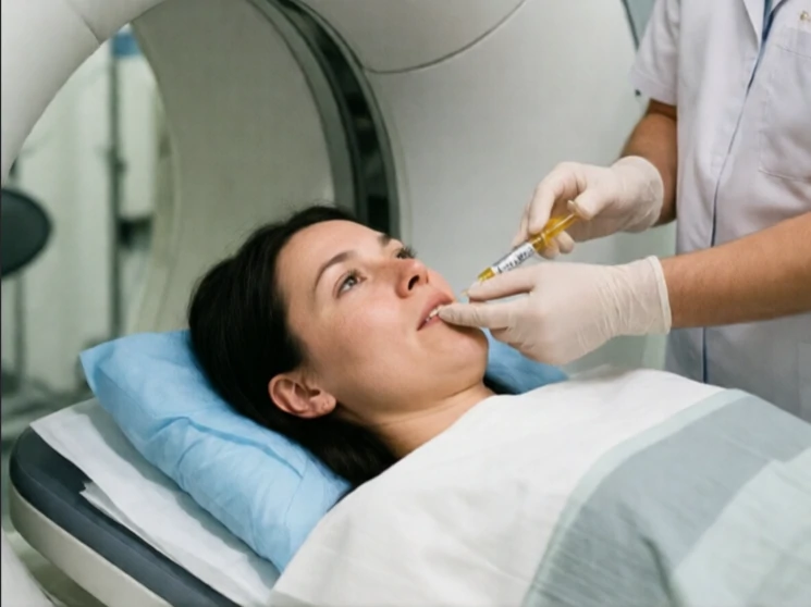
A PET scan, or positron emission tomography scan, produces images of brain activity by detecting the distribution of a radioactive tracer, typically a form of glucose. Regions that consume more glucose appear more active.
The participant receives a small injection of the tracer and then performs a task or rests while detectors surrounding the head record the emitted positrons. A computer reconstructs the data into images that show relative metabolic activity. PET has been used to study the neural correlates of language, memory, emotion, and various psychiatric and neurological disorders. It can also track the distribution of specific neurotransmitter receptors when appropriate labeled compounds are used.
Spatial resolution is lower than that of MRI, and the use of radioactive material limits the frequency of scanning. For many research purposes functional MRI has largely replaced PET because it requires no injection and can be repeated more freely. PET remains valuable for certain clinical questions and for studying molecular targets that MRI cannot address.
When the question is which brain regions are metabolically active during a particular state or task, PET provides a direct answer.
Comments
No comments yet. Be the first to share a thought.
Leave a comment