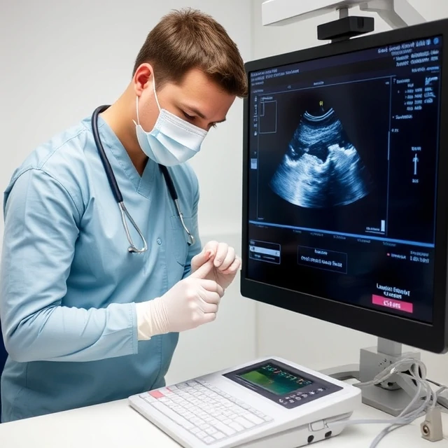
Sound waves above human hearing can paint living anatomy in real time. Ultrasound is an imaging technique that uses high-frequency sound waves to produce pictures of internal organs, vessels, and the developing fetus without ionizing radiation.
A transducer sends and receives echoes; computers convert them into images. Ultrasound excels at examining soft tissues, blood flow (Doppler), and guiding procedures such as biopsies and line placement. It is portable, relatively inexpensive, and safe in pregnancy.
Image quality depends on operator skill and patient factors such as body habitus. Air and bone block the beam, limiting some applications. Point-of-care ultrasound has expanded use beyond radiology departments into emergency, critical-care, and primary-care settings.
- Imaging with high-frequency sound waves
- Safe, portable, and free of ionizing radiation
- Ideal for soft tissue, vessels, and obstetrics
- Operator-dependent and limited by air or bone
Comments
No comments yet. Be the first to share a thought.
Leave a comment