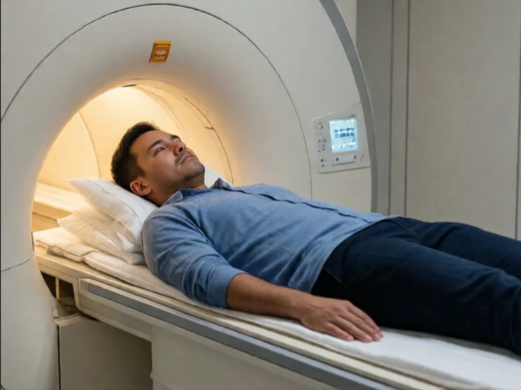
Functional magnetic resonance imaging, or fMRI, measures brain activity by detecting changes in blood flow. When a region of the brain becomes more active, it receives a rapid increase in oxygenated blood; fMRI records that hemodynamic response.
The method has transformed cognitive neuroscience. Researchers can observe which areas are engaged during perception, memory, language, decision making, and emotion while the participant lies in the scanner performing carefully designed tasks. The spatial resolution is on the order of millimeters; the temporal resolution is limited by the slower blood-flow response, typically a few seconds. Combining fMRI with other techniques such as EEG can help compensate for the temporal limitation.
Interpretation requires caution. The BOLD signal is an indirect measure of neural activity, and a “lit-up” region is not necessarily the sole or primary cause of the behavior under study. Careful experimental design, appropriate statistical thresholds, and replication remain essential. Despite these constraints, fMRI has mapped large-scale networks and generated hypotheses that guide both basic research and clinical investigation.
When the question is which parts of the brain are working harder during a particular mental act, fMRI supplies the most widely used answer.
Comments
No comments yet. Be the first to share a thought.
Leave a comment