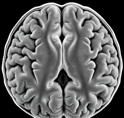
Freshly cut brain tissue reveals two distinct colors. The gray matter consists mainly of neuronal cell bodies, dendrites, unmyelinated axons, and the synapses among them. It forms the outer cortical sheet and also appears in deep nuclei and in the central core of the spinal cord.
Because synapses are concentrated here, gray matter is the primary site of neural computation. Cortical gray matter is organized into layers that differ in cell types and connections. The thickness and integrity of gray matter change across the lifespan and are altered in many neurological and psychiatric conditions; modern imaging quantifies these changes in living people.
In contrast, white matter contains the long-range myelinated axons that link distant gray-matter regions. Both compartments are essential, yet the gray matter is where most local processing and plasticity occur. Histological stains that highlight cell bodies have been used for more than a century to map gray-matter architecture.
Loss of gray matter volume often correlates with cognitive decline in aging and disease.
Comments
No comments yet. Be the first to share a thought.
Leave a comment