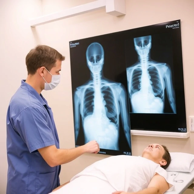
Invisible rays can reveal bone and some soft-tissue shadows. An X-ray is an imaging technique that passes a controlled beam of ionizing radiation through the body onto a detector, producing a two-dimensional projection image of internal structures.
Dense tissues such as bone absorb more radiation and appear white; air-filled spaces appear dark. Plain radiographs remain the first-line study for fractures, pneumonia, and many acute abdominal conditions. Radiation dose is kept as low as reasonably achievable, with special caution in children and pregnancy.
Technologists position patients to obtain standard views; radiologists interpret the images. Portable X-ray units serve intensive-care and emergency patients who cannot travel. Despite the rise of CT and MRI, the plain X-ray is still the most commonly performed medical imaging examination worldwide.
- Projection imaging using ionizing radiation
- Excellent for bone and many acute chest findings
- Dose minimized by technique and shielding
- First-line study for numerous common conditions
Comments
No comments yet. Be the first to share a thought.
Leave a comment