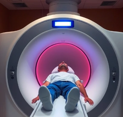
A molecule labeled with a positron-emitting isotope is injected into the bloodstream and accumulates in tissues according to its biological properties. When the emitted positron annihilates with an electron, two gamma rays are produced; detectors surrounding the head register the coincident rays and reconstruct a three-dimensional map of the tracer’s distribution. That is positron emission tomography, or PET.
Depending on the tracer, PET can measure glucose metabolism, blood flow, dopamine-receptor occupancy, amyloid deposition, or many other molecular processes. It therefore provides functional and neurochemical information that structural imaging cannot. Spatial resolution is typically a few millimeters; temporal resolution is limited by the kinetics of the tracer.
PET has been central to the study of neurodegenerative diseases, addiction, and the mechanisms of psychoactive drugs. Combined PET/MRI scanners now allow simultaneous acquisition of molecular and anatomical data. The need for a cyclotron or nearby isotope supply and the use of ionizing radiation are practical constraints.
A well-chosen tracer can make a specific receptor population or pathological protein visible in the living brain.
Comments
No comments yet. Be the first to share a thought.
Leave a comment