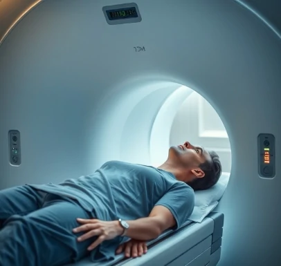
When a brain region becomes more active, local blood flow and oxygenation rise within a few seconds. Functional magnetic resonance imaging detects those hemodynamic changes and thereby maps the location of neural activity noninvasively.
The most common signal is the blood-oxygen-level-dependent, or BOLD, contrast. Participants perform tasks inside the scanner while images are acquired every one to three seconds. Statistical comparison of task and control conditions reveals regions whose activity correlates with the mental operation of interest. The method has transformed cognitive neuroscience by allowing repeated study of the same individuals and by covering the whole brain.
Limitations include relatively slow temporal resolution compared with EEG and an indirect relationship to actual neuronal firing. Careful experimental design and analysis are required to avoid false positives. Despite these constraints, fMRI remains the dominant tool for localizing human brain function in healthy and clinical populations.
A single scanning session can yield both structural images and rich functional datasets.
Comments
No comments yet. Be the first to share a thought.
Leave a comment